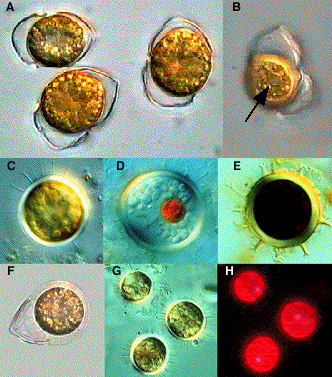
A&B = Cells with split theka (arrow=pyrenoid); C&D = Living cysts; E = Cyst in Lugol; F = Newly formed cyst. (A-F DIC). G&H = Cysts in DIC and in epifl. microscope with blue filter combination (observe white fluorescing body )
Life-form: Solitary
Size: Length 10-35 µm, width 8-29 µm.
Resting spore: +
Note: Dale (1977) as Peridinium faeroense, Dodge (1982) as Scrippsiella faeroense
Distinctive features: Five cingular plates, thecal plates with pores surrounded with concentric rings, Cyst structure
Similar species: Scrippsiella spp.
Distribution:
Literature:
Dale, B. 1977a. New observations on Peridinium faeroense
Paulsen (1905), and classification of small orthoperidinoid
dinoflagellates. Br. Phycol. J. 12: 241-253.
Dodge, J. D. 1982. Marine Dinoflagellates of the British Isles. Her Majesty's Stationery Office, London. 303 pp.
Indelicato, S. R. & Loeblich, A. R., III. 1986. A revision of the marine peridinoid genera (Pyrrophyta) utilizing hypothecal-cingular plate relationships as a taxonomic guideline. Jap. J. Phycol. 34: 153-162.
Lewis, J. 1991. Cyst-theca relationships in Scrippsiella (Dinophyceae) and related orthoperidinoid genera. Botanica Marina. 34: 91-106.